Monday, May 11, 2015

Hyperglycaemic emergencies

Hyperglycaemic emergencies

Diabetic ketoacidosis (DKA) and hyperosmolar hyperglycaemic state (HHS) are diabetes emergencies with overlapping features. With insulin deficiency, hyperglycaemia causes urinary losses of water and electrolytes (sodium, potassium, chloride) and the resultant extracellular fluid volume (ECFV) depletion. (J.Goguen, 2013)

Causes

  • Diabetes mellitus
  • Insulin omission
  • Infection
  • Myocardial infarction
  • Abdominal crisis
  • Trauma
  • Possibly, treatment with insulin infusion pumps, thyrotoxicosis, cocaine, atypical antipsychotics.
  • Possibly, interferon. 

Signs and Symptoms

  • Ketonuria
  • Inability to swallow or keep fluids down
  • Vomiting
  • Persistent diarrhoea
  • Persistently raised glucose (>28 mmol/L) despite increasing insulin.
  • Ketoacidosis is clinically obvious (dehydration, abdominal pain, intractable vomiting, rapid or
    laboured respirations) 

Investigations

  • Elevated plasma and/or urinary ketones

      • Metabolic acidosis (raised H+/low serum bicarbonate)

The presence of the following features should alert you to the possibility of DKA:
      • ketonuria
      • Rapid and deep sighing respirations, smell of ketones
      • Vomiting/abdominal pain
      • Drowsiness/reduced conscious level
      • Intra and extra-vascular volume depletion with reduced skin turgor, tachycardia and hypotension (late features)

  • FBS

  • Blood test to test ketones level in blood serum.

Treatment


  • In the early stages, where patient is fully conscious and able to take adequate oral fluids.
  •  Intravenous insulin, fluids and electrolytes is required to restore the metabolic equilibrium.
  • Administer IV normal saline initially. If the patient is in shock, give 1–2 L/h initially to correct shock; otherwise, give 500 mL/h for 4 hours, then 250 mL/h for 4 hours.
  • Add potassium immediately if patient is normo- or hypokalemic. Otherwise, if initially hyperkalemic, only add potassium once serum potassium falls to <5 to 5.5 mmol/L and patient is diuresing.
  • Once plasma glucose reaches 14.0 mmol/L, add glucose to maintain plasma glucose at 12.0–14.0 mmol/L.
  • After hypotension has been corrected, switch normal saline to half-normal saline (with potassium chloride). However, if plasma osmolality is falling more rapidly than 3 mmol/kg/h and/or the corrected plasma sodium is reduced, maintain IV fluids at higher osmolality (i.e. may need to maintain on normal saline).

REFERENCES


  • Anonymous. (n.d.). Hyperglycaemic Emergencies . Retrieved 05 08, 2015, from www.nhslothian.scot.nhs.uk: http://www.nhslothian.scot.nhs.uk/Services/A-Z/DiabetesService/InformationHealthProfessionals/DiabetesHandbookForPrimaryCare/Hyperglycaemic%20Emergencies.pdf
  • D.Candace, M.Naughton,H.Wesley, S.Corey . (2011, April). Diabetes in the Emergency Department: Acute Care of Diabetes Patients. Retrieved May 08, 2015, from clinical.diabetesjournals.org: http://clinical.diabetesjournals.org/content/29/2/51.full
  • J.Goguen, J. G. (2013). Hyperglycemic Emergencies in Adults. Retrieved 05 07, 2015, from anadian Diabetes Association : http://guidelines.diabetes.ca/browse/Chapter15


Hypoglycemia

Hypoglycemia

Hypoglycemia is characterized by a reduction in plasma glucose concentration to a level that may induce symptoms or signs such as altered mental status and/or sympathetic nervous system stimulation. This condition typically arises from abnormalities in the mechanisms involved in glucose homeostasis. The most common cause of hypoglycemia in patients with diabetes is injecting a shot of insulin and skipping a meal or overdosing insulin. ( O.Hamdy, R.Khardori., 2014)

Causes

1. Side effect of drugs used for the treatment of diabetes
2. Certain medications, such as quinine
3. Drinking too much alcohol
4. Some medical conditions, such as hepatitis or kidney disorders
5. A tumor that produces excess insulin
6. Endocrine disorders, such as adrenal gland deficiency

( https://s-media-cache-ak0.pinimg.com/originals/2b/38/eb/2b38eb9ee3b05e4da4e7046e51c97093.jpg )


Signs and Symptoms of Hypoglycemia

  • Shakiness
  • Nervousness or anxiety
  • Sweating, chills and clamminess
  • Irritability or impatience
  • Confusion, including delirium
  • Rapid/fast heartbeat
  • Lightheadedness or dizziness
  • Hunger and nausea
  • Sleepiness
  • Blurred/impaired vision
  • Tingling or numbness in the lips or tongue
  • Headaches
  • Weakness or fatigue
  • Anger, stubbornness, or sadness
  • Lack of coordination
  • Nightmares or crying out during sleep
  • Seizures 
( http://www.diabetesinfo.org.au/webdata/images/hypoglycemia.gif )

Investigations.

          ·         Vital signs
          ·         Head, eyes, ears, nose, and throat
          ·         Cardiovascular
          ·         Glucose and electrolyte levels (including calcium, magnesium)
          ·         Oral glucose tolerance test and/or 72-hour fasting plasma glucose
          ·         Complete blood count
          ·         Blood cultures
          ·         Urinalysis
          ·         Serum insulin, cortisol levels, and thyroid hormone levels
          ·         C-peptide levels
          ·         Insulin radioimmunoassay
          ·         CT scanning
          ·         MRI
          ·         Octreotide scanning


Treatments


           ·         Glucose supplements (eg, dextrose)
           ·         Glucose-elevating agents (eg, glucagon)
           ·         Inhibitors of insulin secretion (eg, diazoxide, octreotide)
           ·         Antineoplastic agents (eg, streptozocin)
           ·         Fasting hypoglycemia: Dietary therapy (frequent meals/snacks preferred, especially at night, with complex carbohydrates); IV glucose infusion; IV octreotide
           ·         Reactive hypoglycemia: Dietary therapy (restriction of refined carbohydrates, avoidance of simple sugars, increased meal frequency, increased protein and fiber)


REFERENCES


  • O.Hamdy, R.Khardori. (2014, december 22). Hypoglycemia . Retrieved May 7, 2015, from medscape: http://emedicine.medscape.com/article/122122-overview

  • Association, A. D. (2014, September 16). Hypoglycemia (Low Blood Glucose). Retrieved May 7, 2015, from www.diabetes.org: http://www.diabetes.org/living-with-diabetes/treatment-and-care/blood-glucose-control/hypoglycemia-low-blood.html

  • R.Nall. (2014, April 21). Low Blood Sugar (Hypoglycemia). Retrieved May 7, 2015, from www.healthline.com: http://www.healthline.com/health/hypoglycemia#Overview1

Acute Stroke

Acute Stroke

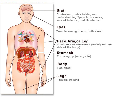
A stroke occurs when the blood supply to your brain is interrupted or reduced. This deprives your brain of oxygen and nutrients, which can cause your brain cells to die.

( http://www.stemcellmd.org/wp-content/uploads/2011/01/si55551195-300x226.jpg )

Causes

  1. A blocked artery (ischemic stroke) - Thrombotic stroke. A thrombotic stroke occurs when a blood clot (thrombus) forms in one of the arteries that supply blood to your brain.

                                                        -  Embolic stroke. An embolic stroke occurs when a blood clot or other debris forms away from your brain,commonly in your heart and is swept through your bloodstream to lodge in narrower brain arteries.

     2. The leaking or bursting of a blood vessel (hemorrhagic stroke) - Hemorrhagic stroke occurs when a blood vessel in your brain leaks or ruptures. There are two types of hemorrhagic stroke including Subarachnoid hemorrhage and Intracerebral hemorrhage.

     3. Temporary disruption of blood flow to their brain (transient ischemic attack, or TIA) - A temporary decrease in blood supply to part of your brain causes TIAs, which often last less than five minutes.
(Myoclinic, 2015)


Signs and symptoms

1. Abrupt onset of hemiparesis, monoparesis, or (rarely) quadriparesis.

2. Hemisensory deficits.

3. Monocular or binocular visual loss.

4. Visual field deficits.

5. Diplopia.

6. Dysarthria.

7. Facial droop.

8. Ataxia.

9. Vertigo (rarely in isolation).

10. Nystagmus.

11. Aphasia.

12.Sudden decrease in level of consciousness. (E.C.Jauch, H.L. Lutsep, 2015)

( http://www.medindia.net/patients/patientinfo/images/stroke.jpg )

Investigations

  • CT angiography and CT perfusion scanning.
  • Magnetic resonance imaging (MRI).
  • Carotid duplex scanning.
  • Digital subtraction angiography.
  • Complete blood count (CBC).
  • Cardiac biomarkers: Important because of the association of cerebral vascular disease and coronary artery disease.
  • Toxicology screening: May assist in identifying intoxicated patients with symptoms/behavior mimicking stroke syndromes.
  • Arterial blood gas analysis: In selected patients with suspected hypoxemia, arterial blood gas defines the severity of hypoxemia and may be used to detect acid-base disturbances.

Treatments


1. Therapy with clot-busting drugs must start within 3 hours if they are given into the vein.

2. Aspirin. Aspirin is an immediate treatment given in the emergency room to reduce the likelihood of having another stroke.

3. Intravenous injection of tissue plasminogen activator (TPA). Some people can benefit from an injection of a recombinant tissue plasminogen activator (TPA), also called alteplase. An injection of TPA is usually given through a vein in the arm. TPA restores blood flow by dissolving the blood clot causing your stroke, and it may help people who have had strokes recover more fully.

4. Medications delivered directly to the brain -  using catheter.

5. Mechanical clot removal.

6. Oxygen - Supplement if indicated (Sa02 < 94%) (T. I. A. ACUTE STROKE MANAGEMENT GUIDELINE ).

( http://www.youtube.com/watch?v=uliThWXvpdg )

REFERENCES

  • E.C.Jauch, H.L. Lutsep. (2015, January 20). Ischemic Stroke . Retrieved May 7, 2015, from medscape: http://emedicine.medscape.com/article/1916852-overview


  • Myoclinic. (2015, april 28). Stroke. Retrieved May 07, 2015, from www.mayoclinic.org: http://www.mayoclinic.org/diseases-conditions/stroke/symptoms-causes/dxc-20117265


Status Epilepticus

Status Epilepticus

Status epilepticus (SE) is a common, life-threatening neurologic disorder that is essentially an acute, prolonged epileptic crisis (see the image below). SE can represent an exacerbation of a preexisting seizure disorder, the initial manifestation of a seizure disorder, or an insult other than a seizure disorder. In patients with known epilepsy, the most common cause is a change in medication. Most seizures terminate spontaneously.  (J.L. Roth, S.A. Berman, , 2014)

Causes

  • Cerebrovascular disorders

  • Brain trauma

  • Infections

  • Low anti-epileptic drug levels in patients with epilepsy.

Signs and symptoms

  • Focal or unilateral paresthesias or numbness.
  • Focal visual changes - usually characterized by flashing lights.
  • Focal visual obscuration or focal colorful hallucinations.
  • Olfactory or gustatory hallucinations.
  • Atypical rising abdominal sensations.
  • Muscle spasms.
  • Loss of bowel or bladder control.
  • Clenched teeth.
  • Irregular breathing.

Investigations

  • EEG
  • Pulse oximetry; blood gases.
  • Blood tests for glucose, renal function, electrolytes, liver function, calcium and magnesium; FBC and clotting; AED levels.
  • 5 ml of serum and 50 ml of urine samples should be saved for future analysis, including toxicology, especially if the cause of the status epilepticus is uncertain.
  • A CT scan or MRI (Tidy, 2012)

Treatments


1. Premonitory stage(pre-hospital) - Diazepam 10−20 mg given rectally, repeated once 15 minutes later. If status continues to threaten, or midazolam 10 mg given buccally.

If seizures continue, treat as below,

2. Early status Lorazepam -  (i.v.) 0.1 mg/kg (usually a 4 mg bolus, repeated once after 10−20 minutes; rate not critical) Give usual AED medication if already on treatment.

For sustained control or if seizures continue, treat as below,

3. Established status - Phenytoin infusion at a dose of 15-18 mg/kg at a rate of 50 mg/minute or fosphenytoin infusion at a dose of 15−20 mg phenytoin equivalents (PE)/kg at a rate of 50-100 mg PE/minute and/or Phenobarbitone bolus of 10-15 mg/kg at a rate of 100 mg/minute.

4. Refractory status - General anaesthesia, with one of: o propofol (1-2mg/kg bolus, then 2-10mg/kg/hour) titrated to effect. o midazolam (0.1-0.2mg/kg bolus, then 0.05-0.5mg/kg/hour) titrated to effect. o thiopentone (3-5mg/kg bolus, then 3-5mg/kg/hour) titrated to effect; after 2-3 days infusion rate needs reduction as fat stores are saturated.

5. Anaesthetic continued for 12−24 hours after the last clinical or electrographic seizure, then dose tapered (giudelines, 2003)


( http://www.educatehealth.ca/media/296429/1-lightbox-epilepsy-treatment-status%20epilepticus.png)

References


  • Guidelines, N. (2003, December). Protocol for treating status epilepticus in adults and. Retrieved April 29, 2015, from www.nice.org.uk: http://www.nice.org.uk/guidance/cg20/documents/appendix-c-protocol-for-status-epilepticus-first-consultation2


  • J.L. Roth, S.A. Berman, . (2014, April 28). Status Epilepticus . Retrieved April 29, 2015, from emedicine.medscape.com: http://emedicine.medscape.com/article/1164462-overview


  • Tidy, C. (2012, 03 14). Status Epilepticus Management. Retrieved 04 29, 2015, from www.patient.co.uk: http://www.patient.co.uk/doctor/status-epilepticus-management


Status Asthmaticus


Status Asthmaticus

Status asthmaticus is a life-threatening form of asthma in which progressively worsening reactive airways are unresponsive to usual appropriate therapy that leads to pulmonary insufficiency. (S. Agarwal, S. Kache.) The primary mechanical event in status asthmaticus is a progressive increase in airflow resistance. Mucous plugging and mucosal edema or inflammation is the major causes for the delayed recovery in status asthmaticus. The combination of hypoxia, hypercapnia, and acidosis, along with the mechanical effects of increased lung volumes may result in cardiovascular depression or cardiovascular arrest. (Ibsen)

Causes 

  • The causes of an acute, severe asthma attack are unknown.
  • Poor control of allergens or asthma triggers .
  • Infrequently use their peak flow meter and inhaled corticosteroids.

Signs and symptoms

  • Cough
  • Dyspnea
  • Wheezing
  • Retractions
  • Prolonged expiratory phase
  • Pulsus paradoxus - systolic blood pressure drop of more than 18 mmHg with inspiration in teenagers or more than 10 mmHg in children.
  • Evidence of cyanosis/hypoxemia - PaO2 less than 60mmHg, change in consciousness.
  • Hypercapnia - PaCO2 greater than 40mmHg in presence of dyspnea and wheezing.
  • Metabolic acidosis.
  • FEV1 or PEFR (peak expiratory flow rate) less than 20% predicted with little or no response to acute therapy
  •  Silent chest
  • Coma

Investigations

1. Vital signs
  • Temperature: fever may indicate URI, pneumonia, other source of infection.
  • Pulse: Usually tachycardia, even before treatment.
  • Respiratory rate: Usually tachypneic.
  • . Blood pressure: Pulsus paradoxus over 10-15 correlates well with moderate to severe disease, as it indicates the effect that air trapping is having on the cardiac output.
2. Breath sounds/Chest exam - I:E ratio is usually 5:2, may be up to 1:4 with a severe attack. The use of accessory respiratory muscles(abdominal paradoxic breathing, sternocleidomastoid use, nasal flaring, intercostal retractions) correlates with the severity of airway obstruction. Feel for the presence of crepitus in the neck or chest wall, signifying air leak and significant obstruction.

3. Cardiac exam

4. Mental status - Confusion or obtundation suggest significant hypercapnia or hypoxemia, and necessitate immediate action.

5. Clinical Asthma Score


6. Pulmonary function tests

7. Chest X-ray

8. Blood gases - Although, blood gases are often discussed in asthma management, they should not be used to determine the need for intubation. The patient’s clinical status should be the grounds for intubation. An attempt at a blood gas often only agitates the patient further exacerbating the reactive airway process.


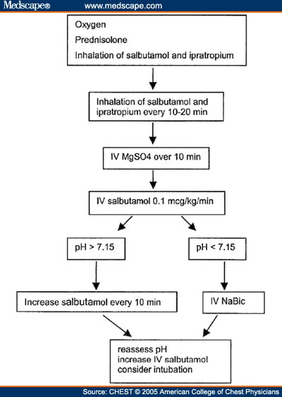
Treatment

1. Intravenous Fluids: Patients in SA are inevitably dehydrated due to poor oral intake, tachypnea, and often emesis. The dehydration often causes a metabolic acidosis as well increasing their work of breathing. Rehydration prevents thickening of mucous secretions and begins to treat the metabolic acidosis.

2. Albuterol: first line therapy
Continuous nebulization - 10-20 mg/hr (or 0.5-0.6 mg/kg/hour) with an oxygen flow rate of 10 – 12L/min.

3. Ipratropium bromide (Atrovent) - 0.25 – 0.5 mg nebulized. Q20 min X 3 doses with continuous albuterol has proven effective in acute management o Every 6- 8 hrs with Albuterol for chronic treatment.

4. Corticosteroids – Solu-medrol - Methyl-prednisone : Loading dose: 2 mg/kg IV o Maintenance dose: 0.5mg/kg IV q 6hr.
( Steroids should be administered IV to SA patients to assure adequate drug delivery in a timely manner. Given the risk of emesis and differential absorption enterally, the drug should not be administered orally.)

5. Magnesium Sulfate - 50 mg/kg IV over 20 min with max of 2 gm.

6. Terbutaline - Loading dose: 5 - 10mcg/kg IV over 10 min o Continuous infusion: 0.4 - 4 mcg/kg/min IV.

7. Mechanical ventilation - Start with low tidal volume, permissive hypercapnia strategy.
                                               1. Tidal volume 4-7 ml/kg (prevents barotrauma / volutrauma, minimize lung distension).
                                                    2. Low Ventilatory rate 10-14 breaths per minute.
                                                    3. I:E ratio 1:4 to 1:6 (avoid air trapping by allowing for complete exhalation).
                                                    4. Tolerate hypercapnia.
                                                    5. Goal pH>7.25 (may require HCO3) .
                                                    6. Peak pressures.
( http://www.youtube.com/watch?v=Px42rbK8fIo )


( http://img.medscape.com/fullsize/migrated/501/434/ch501434.fig1.jpg )

( http://www.samj.org.za/index.php/samj/article/viewFile/6526/5004/34638 )


References


  • Anonymous. (2024). Asthma guide. Retrieved April 28, 2015, from web med: http://www.webmd.boots.com/asthma/guide/status-asthmaticus.


  • Ibsen, L. (n.d.). Status Asthmaticus. Retrieved April 28, 2015, from pedsccm.org: http://pedsccm.org/FILE-CABINET/Practical/Akron_pdfs/7ASTHMAP.PDF.


  • S. Agarwal, S. Kache. (n.d.). STATUS ASTHMATICUS. Retrieved April 28, 2015, from peds.stanford.edu: http://peds.stanford.edu/Rotations/picu/pdfs/14_status_asthmaticus.pdf.


  • S.A.Papiris , E.D.Manali , L. Kolilekas,C. Triantafillidou , I.Tsangaris . (2009). Acute severe asthma: new approaches to assessment and treatment. Retrieved April 28, 2015, from pub med: http://www.ncbi.nlm.nih.gov/pubmed/19911854


Sunday, May 10, 2015

Acute Left Ventricular Failure

Acute Left Ventricular Failure.

Acute left ventricular failure presents as pulmonary oedema due to increased pressure in the pulmonary capillaries. But left ventricular failure and pulmonary oedema are not always synonymous. Acute heart failure may be decompensation of chronic heart failure. (Anonymous, 2006 )

Causes of Acute LVF

  • Acute myocardial infarction or ischemia
  • Aortic stenosis or aortic incompetence
  • Hypertension
  • Mitral incompetence
  • Drugs e.g. beta blockers, cocaine
  • Infection e.g. Myocarditis.
  • Volume overload.
  • Anemia.
  • Hyperthyroidism.

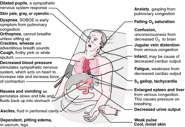
Signs and symptoms

  • Fatigue.
  • Pulmonary oedema.
  • Dyspnoea - Paroxysmal nocturnal dyspnea.
  • Cough.
  • Crepitations - after coughing.
  • Tachycardia.
  • Hypoxia & cyanosis.
  • Sweating.
  • Cardiomegaly - X ray.

  • Dilated pulmonary capillaries and upper lobe diversion - X ray.
  • Gallup rhythm - a third heart sound and/or a fourth heart sound.
  • Orthopnoea.

Signs and symptoms of ALVF
( http://img2.tfd.com/mk/H/X2604-H-14.png )


Investigations

1. ECG

2. Echo cardiograph - The echo cardiograph is the most frequently used investigation. Echo cardiography is the key test to provide a semi-objective assessment of cardiac function. It enables an assessment of:
                        Overall LV systolic function
                        Diastolic function
                        LV wall thickness
                        Valvular diseases.
                        Estimation of pulmonary artery systolic pressure.
3. Serum natriuretic peptides (B‑type natriuretic peptide [BNP] - -  If Acute LVF BNP is more than 100 mg/liter.

4. N‑terminal pro‑B‑type natriuretic peptide [NT‑proBNP] – If acute LVF NT‑proBNP is more than 300 mg/liter.

( In people presenting with new suspected acute heart failure with raised natriuretic peptide levels, perform trans thoracic Doppler 2D echo cardiography to establish the presence or absence of cardiac abnormalities. ) (NICE guidelines, 2014) 

5. Blood tests - FBC, U&E and creatinine, glucose, fasting lipids, thyroid function test
.
6. CXR – Chest X Ray provides details of 
                        Cardiomegaly (cardio thoracic ratio >50%)
                        Ventricular hypertrophy
                        Peribronchial cuffing.
                        Fluid in the fissures
                        Pleural effusions
CXR - ( http://upload.wikimedia.org/wikipedia/commons/1/1c/PulmEdema.PNG )


7. Urinalysis.

8. Lung function tests (peak flow or spirometry).

9. Cardiac magnetic resonance imaging - For assessing ventricular volumes, mass and wall motion. It can be used with contrast to identify inflammation, infiltration and scarring of the myocardium. ( (Kavanagh, 2012).

Management

Initial Management

The patient should be sitting upright and assessed by the ABC approach.

          A -  Check the patient’s airway and administer high flow oxygen through a reservoir bag (also known as trauma mask).

          B - Monitor the patient’s breathing and look for evidence of fatigue (if concerns then an urgent anesthetic/ICU opinion should be sought). Pulse oximetry should be used.

          C - Assess the patient’s circulation by measuring pulse and blood pressure and feeling their peripheries to check perfusion. The patient should be on a cardiac monitor to identify any arrhythmia s. Insert an intravenous cannula.

Immediate management


Morphine IV as required – this will help allay the terror and anxiety but may also reduce preload and after load Oxygen.


Drug therapy

  • Intravenous diuretic (frusemide), venodilator (isosorbide dinitrate), arteriolar dilator (hydralazine), and positive inotropic stimulation (prenalterol) as first-line therapy for acute left ventricular failure.
  • Second line treatments include dobutamine, especially if the systolic blood pressure is below l00mmHg. Bronchodilators such as beta -2 agonists or aminophylline may be used if wheezing is present - 'cardiac asthma'. (Verma SP, Silke B, Hussain M, Nelson GI, Reynolds GW, Richmond A, Taylor SH., 1987) 
1. Diuretics: help by reducing circulatory volume and thereby reducing preload. An intravenous loop diuretic such as Frusemide is administered in the first instance (Frusemide).

2. Venodilators: an intravenous infusion of Glceryl Trinitrate may be useful in reducing preload and after load and may also improve coronary blood flow.

3. Inotropic drugs: these drugs are used to increase myocardial contractility and output. They are often classified according to their activity at alpha and beta receptors.

4. Dobutamine : Exerts its effects on beta 1 and beta 2 receptors and thereby Increases myocardial contractility and output.

5. Dopamine : Exerts its effects on dopaminergic, beta 1 and beta 2 receptors. At low dose (0.5 - 2 mcgs/Kg/min) it has mainly dopaminergic effects ~ increased renal blood flow and diuresis.

6. ACE Inhibitors : ACE inhibitors have been shown to reduce symptoms and signs of heart failure, and improve exercise capacity. Renal function and serum potassium must be monitored before initiation.

7. Spironolactone : Patients with severe heart failure and left ventricular systolic dysfunction should be treated with low dose spironolactone (25mg ) unless there are contra indications. Monitoring of serum potassium is mandatory.

Management of ALVF
( http://eurheartj.oxfordjournals.org/content/ehj/29/19/2388/F7.large.jpg)


Non-pharmacological interventions

1. Diet   
         Salt - Dietary salt should be reduced as much as possible by avoiding the intake of salt rich foods.

         Fluid intake - Excessive fluid intake should be avoided.
         Obesity - Obesity should be reduced. Patients should be advised about setting realistic targets to reduce body weight. This will require counselling about behaviour change as well as nutritional advise. 

 2. Alcohol  - Alcohol should be avoided completely in patients with alcohol induced cardiomyopathy. In other patients with heart failure, alcohol can be consumed in small amounts.

3. Smoking - Patients should be advised to stop smoking and their readiness to do so assessed.

4. Exercise - Appropriate exercise is beneficial for patients with stable heart failure and structured programmes for patients with heart failure, including long term maintenance, are to be developed in the future.

REFERENCES

  • Anonymous. (2006 , august 14). Acute Left Ventricular Failure (LVF). Retrieved April 28, 2015, from www.skills4nurses.com: http://www.skills4nurses.com/index.cgi?article+146

  • Anonymous. (2013). acute left ventricular failure. Retrieved April 28, 2015, from www.gpnotebook.co.uk: http://www.gpnotebook.co.uk/simplepage.cfm?ID=-778436541


  • Kavanagh, S. (2012, 07 29). Heart Failure Diagnosis and Investigation. Retrieved 04 28, 2015, from http://www.patient.co.uk/: http://www.patient.co.uk/doctor/heart-failure-diagnosis-and-investigation


  • Verma SP, Silke B, Hussain M, Nelson GI, Reynolds GW, Richmond A, Taylor SH. (1987, July 10). First-line treatment of left ventricular failure complicating acute myocardial infarction: a randomised evaluation of immediate effects of diuretic, venodilator, arteriodilator, and positive inotropic drugs on left ventricular function. Retrieved april 28, 2015, from pubmed: http://www.ncbi.nlm.nih.gov/pubmed/2441152